Hot NewsLower Leg Bone Diagram Labeled - Femur Definition Function Diagram Facts Britannica
Lower Leg Bone Diagram Labeled - Femur Definition Function Diagram Facts Britannica
Lower Leg Bone Diagram Labeled - Femur Definition Function Diagram Facts Britannica. The bones of the leg and foot form part of the appendicular skeleton that supports the many muscles of the lower limbs. Ebraheim's educational animated video describes the muscle and nerve anatomy of the lower leg.there are fourteen muscles within the lower leg. These muscles work together to produce movements such as standing, walking, running, and jumping. Be able to visualize the skeletal anatomy of the lower leg and hoof of the horse. The fibula (calf bone), the other bone in the lower leg, is connected to the.
Bump at the proximal end of the tibia on the medial side. This keeps the bones together, giving a high ankle sprain time to heal. What is the #1 way to prevent lameness? The knee is the meeting point of the femur (thigh bone) in the upper leg and the tibia (shinbone) in the lower leg. The bones of the leg are the femur, tibia, fibula and patella.
Femur Definition Function Diagram Facts Britannica from cdn.britannica.com Part of the teachme series. The tibia (shin bone) is the medial bone of the leg and is larger than the fibula, with which it is paired (figure 6.52). The medical information on this site is provided as an information. This image is an edited version of this image that was created by user:ladyofhats (mariana ruiz villarreal). The lower leg is also home to nerve fibers. The knee joint is the largest joint in the body and is primarily a hinge joint, although some sliding and rotation occur. The nerves of the leg and foot arise from spinal nerves connected to the spinal cord in the lower back and pelvis. The fibula (calf bone), the other bone in the lower leg, is connected to the.
#1 way to prevent lameness is to purchase a horse with good conformation.
Skeletal muscle anatomy muscular system anatomy human muscular system human muscle anatomy human anatomy and physiology muscle chart anatomy human body systems human body muscles human body organs. Bone diagram forehead (frontal bone) nose bones (nasals) cheek bone (zygoma) upper jaw (maxilla) lower jaw (mandible) breast bone (sternum) upper arm bone (humerus) lower arm bone (ulna) thigh bone (femur) collar bone (clavicle) toe bones (phalanges) ankle bones (tarsals) kneecap (patella) shin bone (tibia) calf bone (fibula) foot bones This area is commonly referred to as the calf. 10 october 2007 (original upload date) The bones of the pelvis and lower back work together to support the body's weight, anchor the abdominal and hip muscles, and protect the delicate vital organs of the vertebral and abdominopelvic cavities. What is the #1 way to prevent lameness? This diagram of a feline skeleton shows you where all of your cat's bones are. The fibula, or calf bone, is smaller and is located on the outside of the lower leg. Part of the teachme series. Any disorder or defect in the knee bone can restrict the activities of the leg which can directly affect our locomotion. Health diagram bone skeleton leg knee science anchor chart human human body. The majority of muscles in the leg are considered long muscles, in that they stretch great distances. Our goal is that these leg anatomy worksheets pictures gallery can be a direction for you, bring you more references and also make you have a great day.
The tibia, also known as the shin bone, is the stronger and larger of the two. The vertebral column of the lower back includes the five lumbar vertebrae, the sacrum, and the coccyx. Lateral view of the bones of the skull unlabeled example. Top flat surface of the tibia on the medial side. The medical information on this site is provided as an information.
Femur Bone Anatomy Labeled Diagram Quiz Color Coded Parts Skeletal System Lower Extremity Ezmed from i.ytimg.com Our goal is that these leg anatomy worksheets pictures gallery can be a direction for you, bring you more references and also make you have a great day. This image is an edited version of this image that was created by user:ladyofhats (mariana ruiz villarreal). Together with the upper leg, it forms the lower extremity. Bump at the proximal end of the tibia on the lateral side. Bones of the pelvis and lower back. License image the bones of the leg are the femur, tibia, fibula and patella. Be able to visualize the skeletal anatomy of the lower leg and hoof of the horse. These muscles work together to produce movements such as standing, walking, running, and jumping.
This image is an edited version of this image that was created by user:ladyofhats (mariana ruiz villarreal).
At the same time, the bones and joints of the leg and foot must be strong enough to support the body. The medial, larger bone of the lower leg. The knee joint is the largest joint in the body and is primarily a hinge joint, although some sliding and rotation occur. Develop an understanding of the causes of equine lameness and methods of treatment. Muscles of the lower limb. Bones of the lower limb. Bone diagram forehead (frontal bone) nose bones (nasals) cheek bone (zygoma) upper jaw (maxilla) lower jaw (mandible) breast bone (sternum) upper arm bone (humerus) lower arm bone (ulna) thigh bone (femur) collar bone (clavicle) toe bones (phalanges) ankle bones (tarsals) kneecap (patella) shin bone (tibia) calf bone (fibula) foot bones Bump at the proximal end of the tibia on the medial side. They support the legs to bear the body weight and also help in proper locomotion. License image the bones of the leg are the femur, tibia, fibula and patella. Includes labels for muscles, bones, nerves and arteries of the knee, leg and foot. Be able to visualize the skeletal anatomy of the lower leg and hoof of the horse. #1 way to prevent lameness is to purchase a horse with good conformation.
The nerves of the leg and foot arise from spinal nerves connected to the spinal cord in the lower back and pelvis. Health diagram bone skeleton leg knee science anchor chart human human body. The anatomical features of the bone are shown on an image with a description to identify the structure and color it on the image. The lumbar plexus forms in the lower back from the merger of spinal nerves l1 through l4 while the. Part of the teachme series.
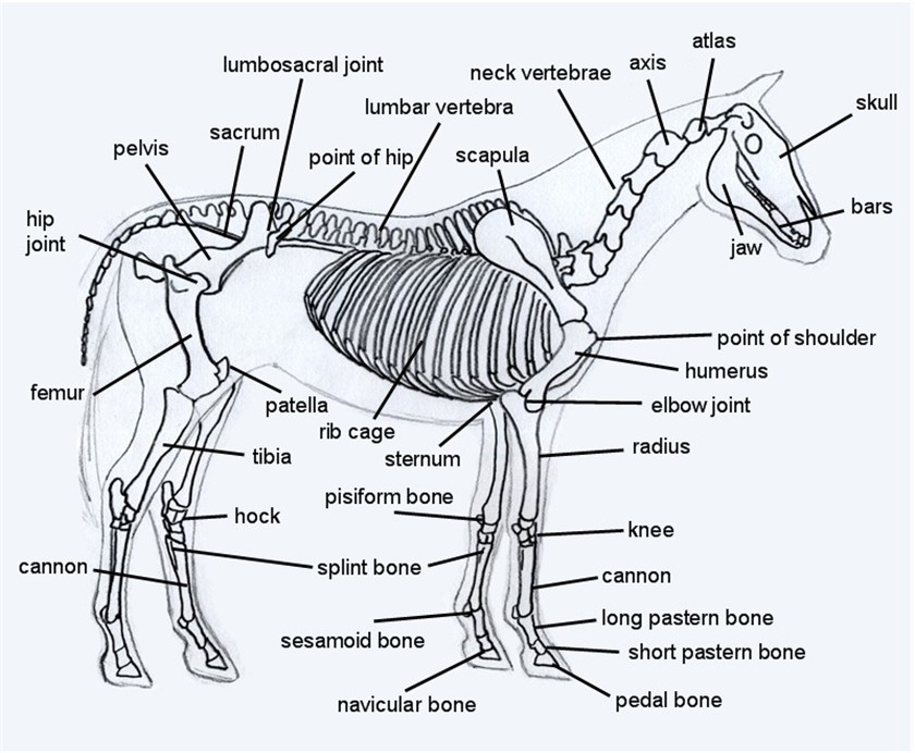
Horse Skeleton Diagram from www.equinespot.com Bones of the lower limb. The knee is the meeting point of the femur (thigh bone) in the upper leg and the tibia (shinbone) in the lower leg. Be able to visualize the skeletal anatomy of the lower leg and hoof of the horse. Nerves of the lower limb. The tibia (also called the shinbone) is located near the midline of the leg. Bones of the leg and foot. Bump at the proximal end of the tibia on the medial side. The fibula, or calf bone, is smaller and is located on the outside of the lower leg.
At the same time, the bones and joints of the leg and foot must be strong enough to support the body.
Together with the upper leg, it forms the lower extremity. Labeled human leg bones created for use in leg bone. 10 october 2007 (original upload date) Be able to visualize the skeletal anatomy of the lower leg and hoof of the horse. Anatomy coloring book coloring books coloring sheets science classroom teaching science classroom ideas science lessons life science science experiments. This area is commonly referred to as the calf. Bones of the lower limb. The bones of the leg are the femur, tibia, fibula and patella. Skull bones unlabeled anatomy bones, skull anatomy, gross anatomy, human. The knee joint is the largest joint in the body and is primarily a hinge joint, although some sliding and rotation occur. It is located toward the middle of the lower leg. The nerves of the leg and foot arise from spinal nerves connected to the spinal cord in the lower back and pelvis. Part of the teachme series.
As these muscles contract and relax, they move skeletal bones to create movement of the body leg bone diagram. Labeled muscles of lower leg.


0 comments